HER 2 Testing
Author Lisa James
Molecular Cytogenetics Service
The Molecular Cytogenetics service within Cellular Pathology based at Heartlands Hospital provides a high quality, fully UKAS accredited FISH service for both internal referrals and external users, both regionally and nationally. Our highly experienced FISH team utilises an integrated approach incorporating morphology, immunohistochemistry (IHC) and fluorescence in-situ hybridisation (FISH) genetics testing to provide a fully comprehensive service for our users. The section currently offers prognostic IHC and FISH HER2 testing for breast and gastric cancers and diagnostic/prognostic FISH testing of lymphomas.
Breast and Gastric Referrals
IHC testing using the 4B5 antibody clone (Roche) is initially performed to screen patient samples for HER2 protein expression levels to identify tumours which “over-express” this protein. Patients with tumours which are HER2 positive usually benefit from treatment with Trastuzumab (Herceptin). Where expression levels are equivocal (2+ IHC staining, see Fig 1.), FISH is carried out to determine unequivocally whether the tumour is positive or negative for HER2 gene amplification (see Fig 2.). Service users may send referrals for both HER2 IHC and FISH testing when appropriate or FISH analysis only if performing their own HER2 IHC.
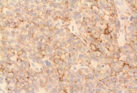
Fig. 1. 2+ IHC staining
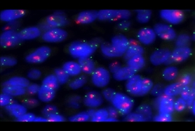
Fig2. HER2 amplified FISH result
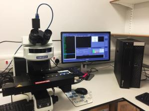
The service performs approximately 4,400 HER2 IHC tests and 1,200 HER2 FISH tests per annum. The turnaround times (TaT) for HER2 breast cancer referrals and gastric referrals is 14 days and 7 days respectively including weekends. However, since the recent introduction of the Abbott Bioview automated digital FISH image capture and analysis system (see Fig 3.) into the department, the TaT for breast and gastric referrals can be quicker than the times stated above.
Lymphoma referrals
We currently offer an extensive repertoire of FISH testing to identify specific gene rearrangements associated with a range of lymphomas/lymphoproliferative disorders in order to aid diagnosis, tumour classification and disease prognosis (see Table 1.). The variety of FISH testing reflects the 2016 revision of the World Health Organization classification of lymphoid neoplasms
| FISH Test | Probe Type | Disease |
| MYC | Dual colour breakapart | Burkitt lymphoma, high grade lymphoma with MYC and BCL2 and or BCL6 translocations, high grade lymphoma NOS, DLBCL |
| IGH/MYC* | Dual colour, dual fusion | |
| MYC/IGK/IGL* | Tri-colour, dual fusion | |
| MYC/BCL6* | Dual colour, breakapart | |
| BCL2 | Dual colour, breakapart | High grade lymphoma with MYC and BCL2 and/or BCL6 translocations, folicular Lymphoma |
| BCL6 | Dual colour, breakapart | High grade lymphoma with MYC and BCL2 and/or BCL6 translocations, folicular Lymphoma |
| MALT1 | Dual colour, breakapart | MALT1 lymphoma |
| TP53 | Dual colour, site specific | CLL |
| ATM | Dual colour, site specific | CLL |
| IRF4/DUSP22* | Dual colour, breakapart | Anaplastic Large Cell Lymphoma (ALCL), large B-cell lymphoma |
|
COND1 |
Dual colour, breakapart | Mantle cell lymphoma |
| IGH | Dual colour, breakapart | General FISH marker for all lymphomas |
Table 1. List of lymphoma/LPD FISH probes currently offered by the molecular cytogenetics service.
The FISH testing strategy (see Fig 4.) to exclude a high grade lymphoma with MYC and BCL2 and/or BCL6 translocations involves initial testing using a MYC breakapart probe.
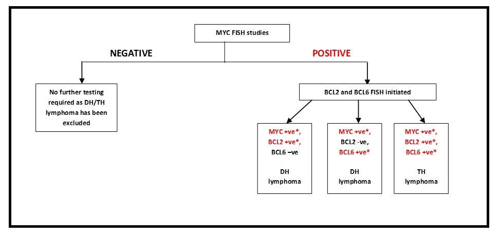
Fig. 4. Flowchart of current high grade lymphoma FISH testing strategy, DH= double-hit, TH = triple-hit *(Includes non-classical abnormal signal patterns)
Additional BCL2 and BCL6 FISH testing will only be performed if the MYC FISH test is abnormal unless specifically requested (see Fig 5.).
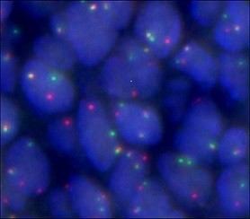
Fig 5. Abnormal 1F1R1G split signal pattern using the Vysis MYC dual colour, breakapart probe specific for the MYC gene region at 8q24. BCL2 and BCL6 FISH studies will be carried out (see Fig 6).
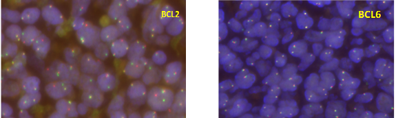
Fig. 6 Vysis dual colour, breakapart BCL2 and BCL6 FISH results. The BCL2 probe shows an abnormal 1F1R1G signal pattern consistent with a BCL2 gene rearrangement at 18q21. The BCL6 probe shows a normal 2F signal pattern. As the MYC gene is also rearranged this result is consistent with a High grade lymphoma with MYC and BCL2 translocations.
Turnaround Times (TaT’s) for FISH testing
- The TaT for HER2 IHC is 1-2 days from receipt of the sample.
- The TaT for breast HER2 FISH testing is 14 days from receipt of the sample.
- The TaT for gastric HER2 and MYC FISH testing is 7 days from receipt of the sample
Sample Requirements
All samples submitted for FISH testing must be accompanied by a request form detailing the patient name, DOB, NHS number and laboratory number of the requesting laboratory. A copy of the histology report should also be included.
A representative paraffin wax block should be submitted for testing, which should contain sufficient tumour material for FISH testing. Alternatively, we do accept tissue sections on charged, Menzel Glazer slides.
If slides are to be sent please provide 5 sequential sections, cut at 3-4μm, on to charged slides, maintaining placement and orientation on the slide. The slides must be labelled with the laboratory number, including any suffix/part number, patient name and the date that the sections were cut. Sections must be dried overnight at 37OC. Do not heat on.
If slides are being sent for HER2 FISH analysis only, please ensure that the original HER2 immunohistochemistry slide is also included.
Please ensure that spare sections being sent are representative of the IHC slide.
Limitations of test
Samples should preferably be fixed in 10% neutral buffered formalin for 6 – 48 hrs. Suboptimal fixation, either insufficient or extended, can cause issues with interpretation of FISH results.
The use of marker dyes, such as eosin and carbol fuchsin, on breast core biopsies and resection specimens should be avoided as this causes auto-fluorescence on FISH samples which are difficult to interpret.
FISH studies involving bone marrow trephines where tissue decalcification has occurred may fail due to destruction of cellular DNA.
For lymphoma FFPE testing, FISH results are not diagnostic and should be considered in the context of the morphology and immunophenotype. FISH false positive signal patterns may be seen in up to 5% of cells for breakapart probes consequently abnormal clones below these levels may not be excluded.
Contact Details For Molecular Cytogentetics (FISH) Service
Hours of Service: The HER2 service is staffed Monday – Friday 08:30 – 17:00
Please contact our team via our generic email address, This email address is being protected from spambots. You need JavaScript enabled to view it. or telephone the Cellular Pathology secretaries on 0121 4243487 who will transfer you to the FISH service. Alternatively, contact our senior scientist team individually via the following email addresses;
- Dr. Jane Starczynski – Consultant Clinical Scientist This email address is being protected from spambots. You need JavaScript enabled to view it.
- James Dowds - Senior BMS This email address is being protected from spambots. You need JavaScript enabled to view it.
- Julie Pritchard – Senior BMS This email address is being protected from spambots. You need JavaScript enabled to view it.
For Clinical advice and interpretation please contact one of the pathologists listed below.
- Dr N. Dalal, Departmental Consultant Histopathologist (Breast)
- Dr B. Tanchel Consultant Histopathologist (Breast)
- Dr Z. Rudzki Consultant Histopathologist (Lymphoma)
- Dr S Karim Consultant Histopathologist (Breast)
- Dr Y Maurice, Consultant Histopathologist(Breast)
- Dr G Langman, Consultant Histopathologist (Gastric)
Additional Information
- Referred blocks are returned to the referring laboratory by recorded delivery once the case has been reported.
- HER2 IHC and FISH tests are fully FDA approved and validated assays (Pathway 4B5 antibody from Roche and PathVysion dual colour, site specific HER2 FISH probe from Abbott Molecular (both CE marked)).
- The Bioview automated system has been fully validated and is in scope of ISO 15189 UKAS accreditation.
- All FISH and IHC testing is performed in accordance with National Guidelines. We are a national reference centre laboratory for HER2 testing.
- We actively participate in national UKNEQAS/GENQA schemes for HER2 and lymphoma/LPD FISH testing.
- We are continually monitoring and auditing all aspects of our service to provide high quality results for our users.
- Created on .
- Last updated on .